
Figure.
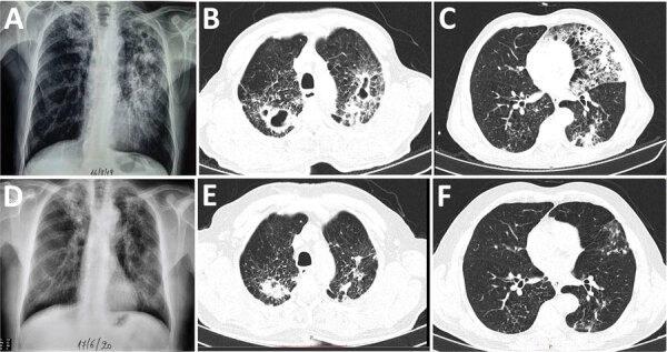
Comparison of chest radiographs and computed tomography scans before and after the end of treatment for a 65-year-old male goat breeder infected with Mycobacterium caprae, northern Greece, 2019. Multiple cavity infiltrations and opacities are shown on the chest radiographs (A) and the computed tomography scan (B, C), mainly in the left upper lobe. After treatment, significant improvement is shown by cavity closure and recession of opacities and infiltration on the chest radiograph (D) and on the chest computed tomography scans (E, F).