
Figure 1.
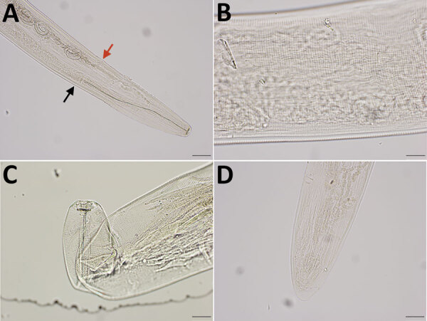
Integrated diagnostic approach for confirming Thelazia callipaeda nematodes: morphologic identification. Specimens were cleared in lactophenol before examination under an Olympus compound microscope (BX53) (https://www.olympus-lifescience.com). Images were taken with an Olympus DP73 camera, and morphometry was performed by using Olympus cellSens software. A) Cephalic end of a female worm. Black arrow indicates esophageal intestinal junction; red arrow indicates vulval opening. Original magnification ×100. B) Transverse striations (150–190/mm) in the cuticle of midbody region of a female worm. Original magnification ×200. C) Buccal cavity of a female worm, wider than deep. Note tightly spaced cuticular striations in the cephalic end. Original magnification ×200. D) Caudal end of female worm with protruding phasmids in the tip. The tail was not protruding unilaterally. Original magnification ×100.