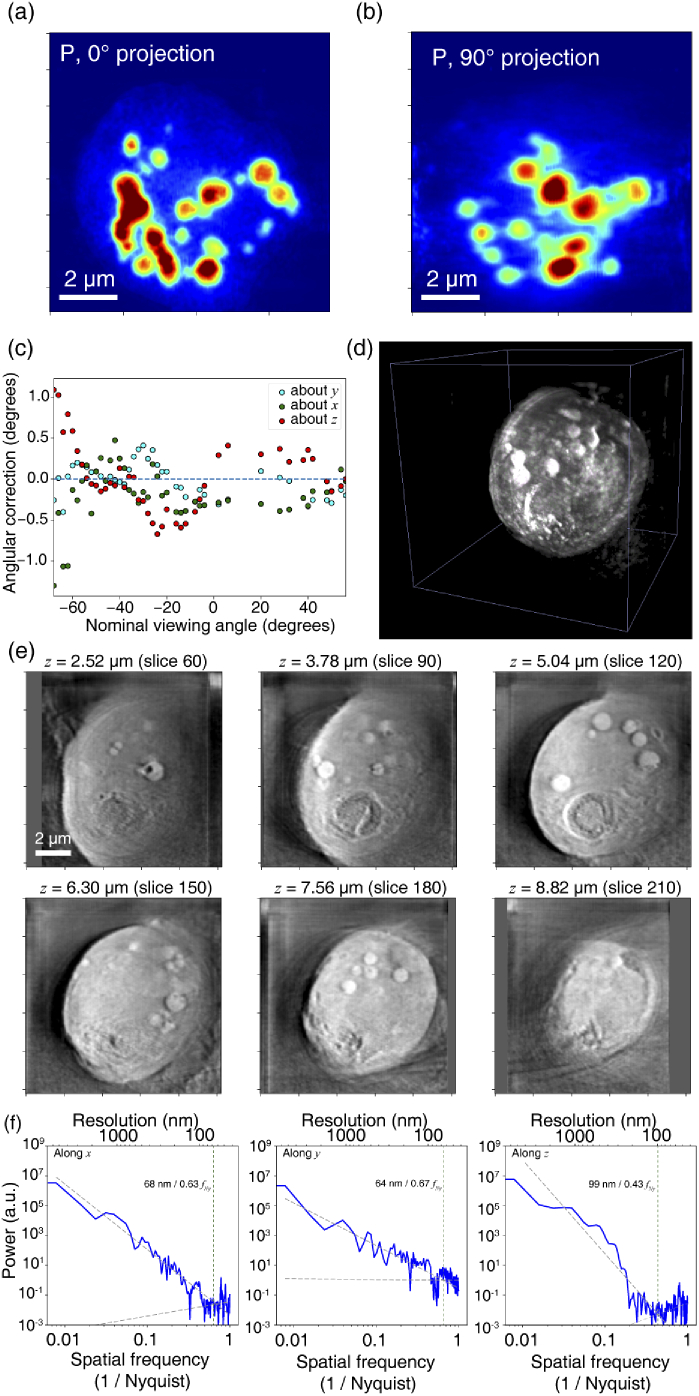
Fig. 12.
Adorym reconstruction of a tomographic X-ray fluorescence and ptychography dataset of a frozen hydrated alga cell Chlamydomonas reinhardtii acquired using 5.5 keV X rays [77]. The phosphorus fluorescence projection (a) at through the 3D volume highlights polyphosphate bodies, which are also seen at somewhat lower resolution in the projection through the reconstruction due to the limited tomographic tilt range of to . As in a previously-reported reconstruction of this dataset [77], this phosphorus reconstruction used refinement of the tomographic tilt angles, leading to angular corrections about all three axes as shown in (c). These refined angles were then employed in a subsequent ptychotomography reconstruction, yielding a 3D volume rendered in (d). Sub-images in (e) show optical sections from the reconstructed volume cut at indicated -positions after it is rotated about the -axis by . In (f), the power spectra of the reconstructions in the -, -, and -direction are shown. For each case, two lines with the same color are fitted respectively using datapoints with spatial frequency in the range of 0.008–0.31 and 0.47–1.0. As the latter fits the noise plateau, the intersection between both lines (marked by dotted vertical lines) provides a measure of spatial resolution.

