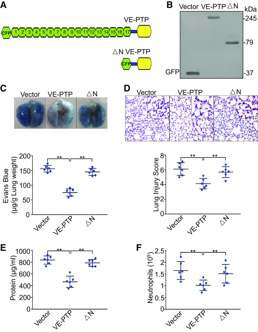
Figure 7.
Exogenous expression of VE-PTP in pulmonary vasculature rescues YAP deficiency-mediated lung injury and inflammation. Endothelial cell-specific YAP knockout mice (YAP-CKO) overexpressing full-length VE-PTP and VE-PTP mutant (△N) were ventilated with a tidal volume of 21 mL/kg for 4 h. A: schematic representation of full length VE-PTP and △N VE-PTP (lacking extracellular domain) mutant. B: verification of overexpression of VE-PTP and VE-PTP mutant (△N) in lung endothelial cells of endothelial cell-specific YAP knockout mice (YAP-CKO) by Western blot. Green fluorescent protein (GFP) for detection of cyan fluorescent protein-tagged VE-PTP and VE-PTP mutant (△N). C: pulmonary vascular permeability measured by Evans blue dye extravasation. Top: representative lung appearance after Evans blue dye administration. Bottom: quantitative analysis of Evans blue-labeled albumin extravasation. n = 6/group (3 male and 3 female mice). **P < 0.001, one-way ANOVA with Bonferroni post hoc test. D: hematoxylin and eosin staining of lung sections. Top: representative lung histology. Magnification, ×20; inset, ×40. Bottom: quantification of histopathological lung injury scores. n = 6/group (3 male and 3 female mice). **P < 0.001, one-way ANOVA with Bonferroni post hoc test. All images for control and treatment groups were collected at the same time under the same conditions. E: protein concentrations in bronchoalveolar lavage fluid. F: neutrophil count in bronchoalveolar lavage fluid by cytospin analysis. n = 6/group (3 male and 3 female mice). **P < 0.001, one-way ANOVA with Bonferroni post hoc test. Data represent means ± SD. VE-PTP, vascular endothelial protein tyrosine phosphatase; YAP, Yes-associated protein.

