Figure 6.
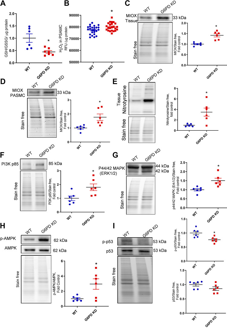
G6PD deficiency-induced oxidative stress and proliferative signaling. G6PD deficiency attenuated GSH/GSSG ratios (A). Isolated pulmonary artery smooth muscle cells (PASMCs) showed increased production of reactive oxygen species (H2O2) than WT cells (B). G6PD KD lung tissue showed increased expression of myo-inositol oxygenase (MIOX) in lung tissue (C) and PASMCs (D). Elevated oxidative stress produced increased reactive nitrogen species (RNS), which was identified by increased expression of global tyrosine nitration in G6PD KD lung tissue (E). Increased ROS/RNS upregulated PI3K p85, ERK1/2, and AMPK pathways (F–H) in G6PD KD lung tissue. Proapoptotic protein p53 phosphorylation was significantly attenuated in G6PD KD lung tissue (I). Data are represented as means ± SE normalized on the total protein (stain-free), n = 6, *P < 0.05 vs. control by unpaired t test or Mann–Whitney U t test. G6PD KD, glucose-6-phosphate dehydrogenase knockdown; GSH, glutathione; GSSG, oxidized glutathione; ROS/RNS, reactive oxygen species/reactive nitrogen species; WT, wild-type.

