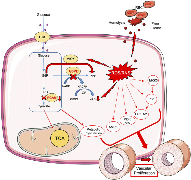
Figure 7.
Schematic diagram depicting the role of G6PD deficiency in vascular remodeling. G6PD deficiency in erythrocytes induced increased hemolysis and free hemoglobin release. Increased free heme in circulation upregulated oxidative stress and barrier dysfunction. Cellular glucose uptake is increased in the G6PD KD lungs. This results in increased glycolytic metabolism; however, the inhibition of PGAM redirected glycolysis towards a nonoxidative branch of PPP and fatty acid synthesis. Increased MIOX pathway, together with decreased GSH production, results in increased ROS/RNS generation. Thus, increased hemolysis and oxidative stress collectively contributed to the upregulation of proliferative signaling p38 MAPK, ERK1/2, PI3K, and AMPK pathways. This leads to vascular proliferation and remodeling. In summary, G6PD deficiency-induced metabolic dysfunction, oxidative stress, and vascular proliferation are the causes of PH pathogenesis in the G6PD KD model. Figure 7 was prepared using templates from the image bank of Servier Medical Art (http://smart.servier.com/) under CC-BY 3.0 license. G6PD KD, glucose-6-phosphate dehydrogenase knockdown; GSH, glutathione; MIOX, myo-inositol oxygenase; p3 MAPK, p38 mitogen-activated protein kinase; PGAM, phosphoglycerate mutase; PPP, pentose phosphate pathway; ROS/RNS, reactive oxygen species/reactive nitrogen species.

