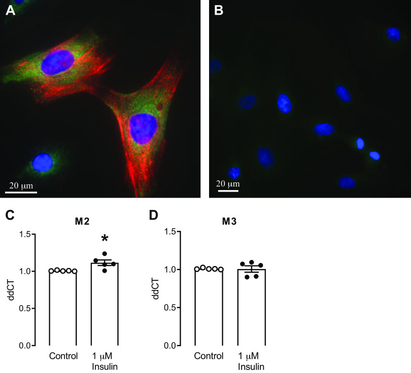
Figure 4.
Rat tracheal smooth muscle cells isolated from wild type rats. Cell phenotype was verified by staining with anti-actin antibody (A; red). Insulin receptor expression on these tracheal smooth muscle cells is shown by positive staining with an anti-insulin receptor β antibody (A; green). B: no primary control for both antibodies; (blue, DAPI nuclear stain). Insulin (1 μM for 3 h) increased M2 muscarinic receptor mRNA (C), but did not change M3 muscarinic receptor mRNA expression (D) in cultured rat tracheal smooth muscle cells. Data shown are means ± SE. *P ≤ 0.05 (n = 5 cultures from separate rats).

