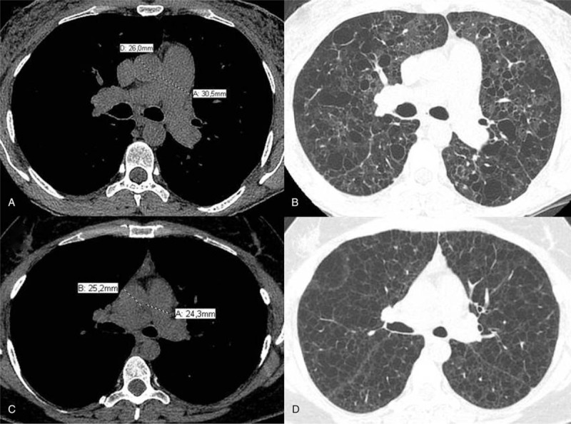
Figure 1.

(A) Axial computed tomography slice at the level of bifurcation of the main pulmonary artery demonstrates measurements of the diameters of the main pulmonary artery (30.5 mm) and ascending aorta (26 mm) in a patient with pulmonary Langerhans cell histiocytosis. PA/Ao ratio in this patient was 1.17. (B) Axial computed tomography image shows irregular pulmonary cysts in the patient presented in (A). (C) Axial computed tomography slice at the level of bifurcation of the main pulmonary artery demonstrates measurements of the diameters of the main pulmonary artery (24.3 mm) and ascending aorta (25.2 mm) in a patient with lymphangioleiomyomatosis. PA/Ao ratio in this patient was 0.96. (D) Axial computed tomography image shows extensive and diffuse pulmonary cysts with regular walls in the patient presented in (C). Ao = ascending aorta, PA = pulmonary artery.
