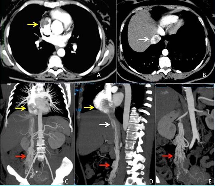
Figure 1 .

CT scan images (A–B) arterious phase showing the defect inside right atrium (yellow arrow) and intrahepatic inferior vena cava (white arrow) due to the presence of the intravascular leimyoma. (C–D) vascular CT reconstruction of the ovarian veins (red arrow), inferior vena cava (white arrow) and right atrium (yellow arrow) stored with the intravascular leiomyoma. (E) Particular view of the ovarian veins (red arrow) with the origin from the right uterine aspect.
