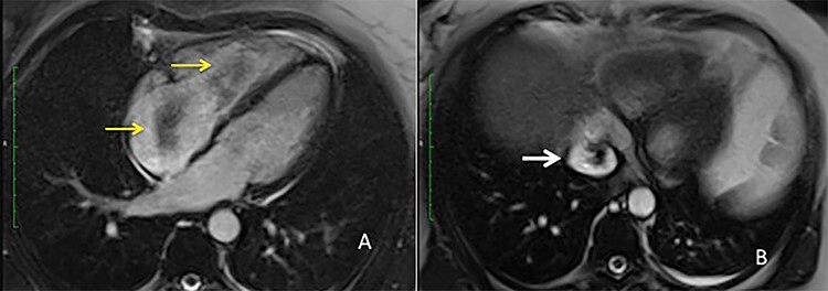
Figure 2 .

MRI view (A) right atrium with the intravascular leiomyoma prolapsing into the right ventricle (yellow arrow) (B) intrahepatic inferior vena cava stored with the intravascular leiomyoma (white arrow).

MRI view (A) right atrium with the intravascular leiomyoma prolapsing into the right ventricle (yellow arrow) (B) intrahepatic inferior vena cava stored with the intravascular leiomyoma (white arrow).