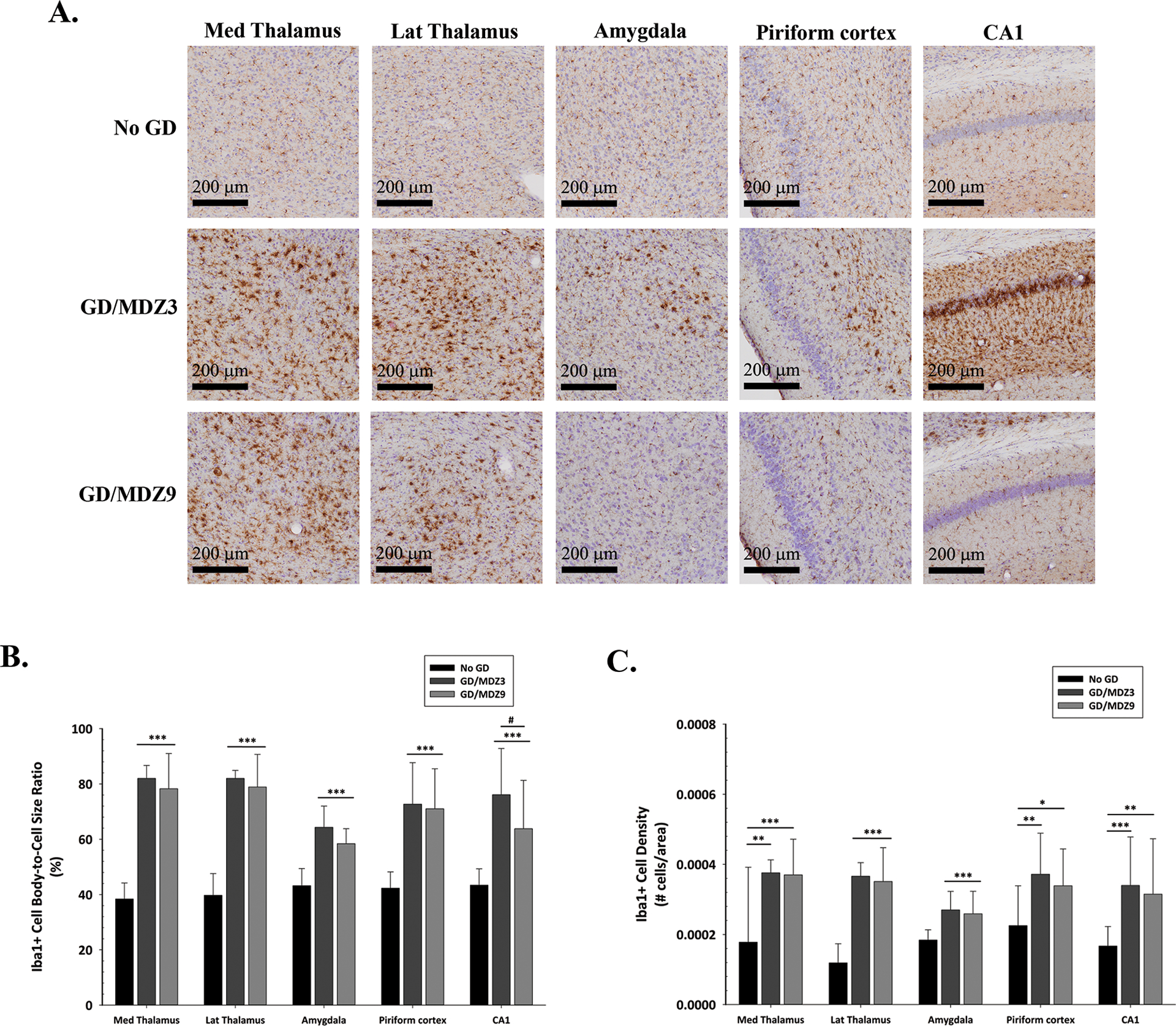
Figure 7.

Midazolam (MDZ) treatment delayed to 40 min after GD-induced seizure failed to prevent microglial activation in ES1−/− mice, independent of sex. (A) Representative images of Iba1-immunostained brain samples from mice exposed SC to GD (82 μg/kg) that received 3 mg/kg MDZ (GD/MDZ3; n = 10) or 9 mg/kg (GD/MDZ9; n = 11) of MDZ 40 min after seizure onset, compared with control (No GD; n = 14) mice. (B) The average (± SD) cell-body-to-cell-size ratios and (C) density of ionized calcium–binding adaptor molecule 1 (Iba1), a marker for microglia, are shown for the dorsomedial thalamus (Med Thalamus), dorsolateral thalamus (Lat Thalamus), basolateral amygdala (Amygdala), layer 3 of the piriform cortex, and the CA1 of the hippocampus. Cresyl violet (purple) counterstain was used for visualization of anatomic landmarks. *P < 0.05, **P < 0.01, ***P < 0.001, compared with the No GD group. #P < 0.05, GD/MDZ3 compared with the GD/MDZ9 group.
