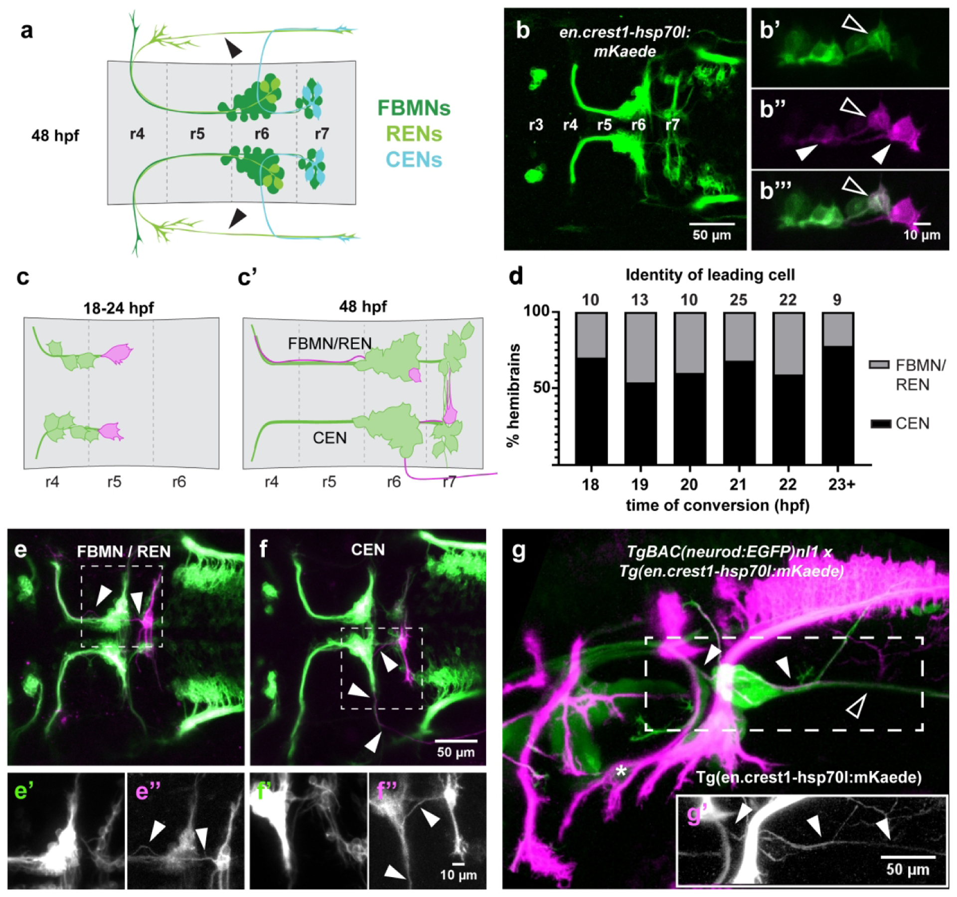
Figure 1: Single-cell photoconversions reveal that OENs migrate concurrently with FBMNs.

(a) Schematic of FBMN, REN, and CEN somas and axon morphologies at 48 hpf. The REN projection crossing the otic vesicle (arrowheads) is not visible in all specimens. (b) The newly-generated Tg(en.crest1-hsp70l:mKaede) line uses the islet1 zCREST1 enhancer (Uemura et al., 2005) to drive expression of the photoconvertible protein Kaede in a subset of cranial efferent neurons. (b’-b”’) Single-cell labeling via photoconversion of Kaede from green to red. A fully converted leading cell and its trailing axon (closed arrowheads) are identified by the presence of red and absence of green protein. Contrast with partial conversion of a follower, in which green protein remains (open arrowheads). (c) Schematic of photoconversion experiments targeting leading mKaede-expressing neurons between 18–24 hpf and (c’) screening criteria for axon morphology of different cell types at 48 hpf. (d) CENs are present in the leading position in over 50% of embryos at every time point between 18–24 hpf. (e) Axon morphology typical of FBMN/RENs (arrowheads); (e’-e”) insets of boxed area show separated green and red channels, respectively. (f) Axon morphology typical of CENs (arrowheads); (f’-f”) insets of boxed area show separated green and red channels, respectively. (g-g’) Double transgenic line with converted Tg(en.crest1-hsp70l:mKaede) and Tg-BAC(neurod:EGFP)nl1 demonstrates that efferent projections leaving r6 (red; closed arrowheads) fasciculate with the sensory afferent projections to the lateral line (green; open arrowheads). Glossopharyngeal motor neurons (nIX) are indicated by an asterisk.
