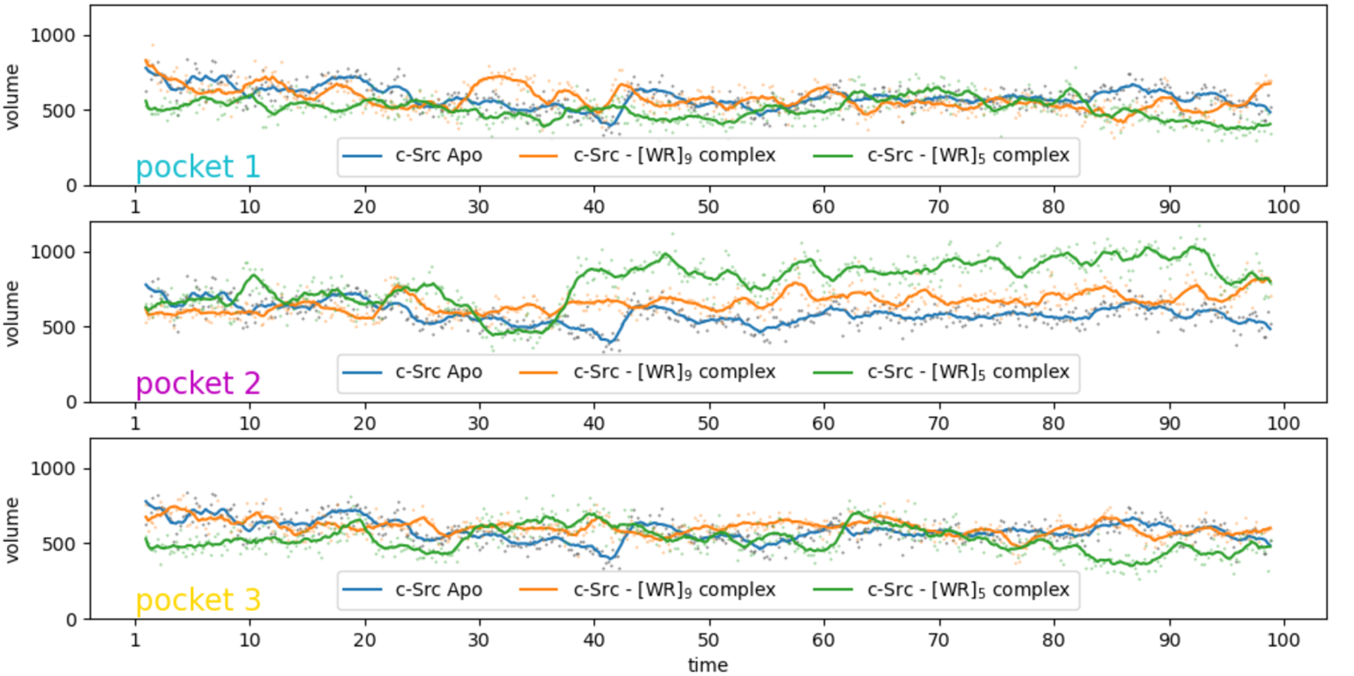
Figure 6.

Volume of the ATP-binding pocket during the MD simulations of the c-Src Apo structure (blue) and with [WR]5 and [WR]9 bound to c-Src (green and gold, respectively) in the 3 identified binding pockets. The volume values are depicted as dots and the lines provide a moving average using a window of 10 points. The peptide bound in pocket 2 modulates the volume of the ATP binding pocket.
