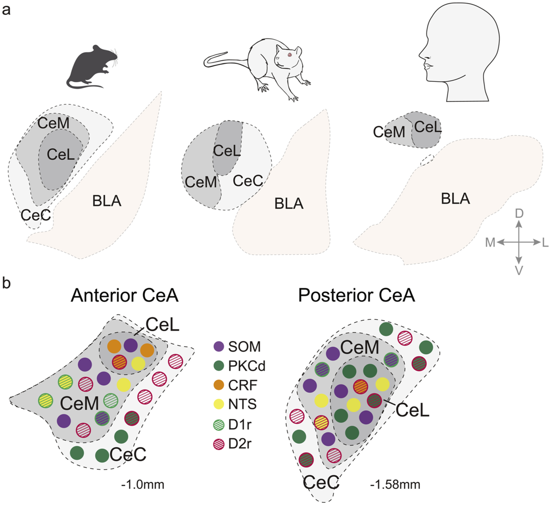
Fig. 2. Species-specific CeA Anatomy and Cell-type distribution.

(a) Coronal views show central nucleus of amygdala and basolateral amygdala organization in mouse (left), rat (middle), or human (right) (adapted from [166–168]. (b) Coronal views show CeA neuronal subtypes and their approximate anatomical distribution across sub-regions in anterior CeA and in posterior CeA, based on expression of select mRNA markers in mice [40, 46]. This figure does not reflect the fact that some neuronal subtypes co-express multiple neurotransmitter peptides (e.g., NTS cells also express SOM), nor is it meant to depict exact proportions of specific cell types. Abbreviations: CeC, central CeA; CeM, medial CeA; CeL, lateral CeA; BLA, basolateral amygdala; D, dorsal; V, ventral; M, medial; L, lateral; A, anterior; P, posterior; SOM, somatostatin; PKCd, protein kinase-C delta; CRF, corticotropin releasing factor; NTS, neurotensin; D1r, dopamine receptor type 1; D2r, dopamine receptor type 2.
