Figure 1.
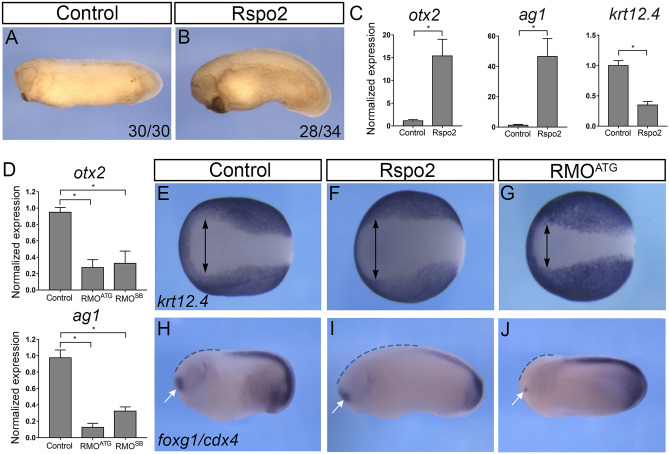
Rspo2 function is essential for anterior development. (A,B) Four-cell embryos were injected with 0.5 ng of Rspo2 RNA into both animal-ventral blastomeres and cultured until stage 28. (A) Uninjected control embryo. (B) Representative embryo injected with Rspo2 RNA. The penetrance is indicated as the ratio of the number of embryos with the phenotype and the total number of injected embryos. (C) The effect of Rspo2 on gene marker expression. Animal pole explants were dissected at stage 9 from embryos overexpressing Rspo2 RNA or uninjected controls. RT-qPCR analysis was carried out for otx2, ag1, and krt12.4 at stage 18. (D) Altered gene expression in Rspo2 morphants. RNA was isolated from stage 18 control embryos or embryos depleted of Rspo2. RT-qPCR for ag1 and otx2 was carried out in triplicates. (C,D) Each graph is a single experiment with triplicate samples, representative from at least 3 independent experiments. Means + /− s. d. are shown. Statistical significance has been assessed by Student’s t test, *, p < 0.05. (E–J) In situ hybridization of control and manipulated stage 16 or 25 embryos with krt12.4 (E–G), foxg1 and cdx4 (H–J) probes. (E–G) Width of the anterior neural plate is shown as lack of krt12.4 (arrows). (H–J) The foxg1 domain is indicated by white arrows, the anterior region lacking cdx4—by dashed lines. See Supplementary Table 1 for quantification.

