
Fig. 3.
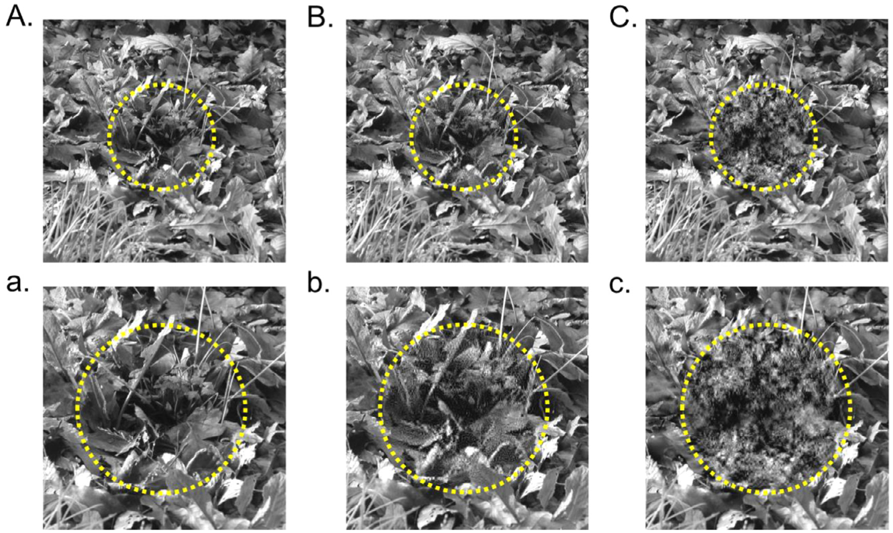
Using wavelets to degrade a spatially-restricted area. (A) Intact natural image with dashed circle denoting the targeted foveal region. (B) Natural image with only fine scale structure degraded near the fovea. (C) Natural image with all structure degraded near the fovea. Lowercase a-c show zoomed-in views of the central regions in A-C.