
Figure 2.
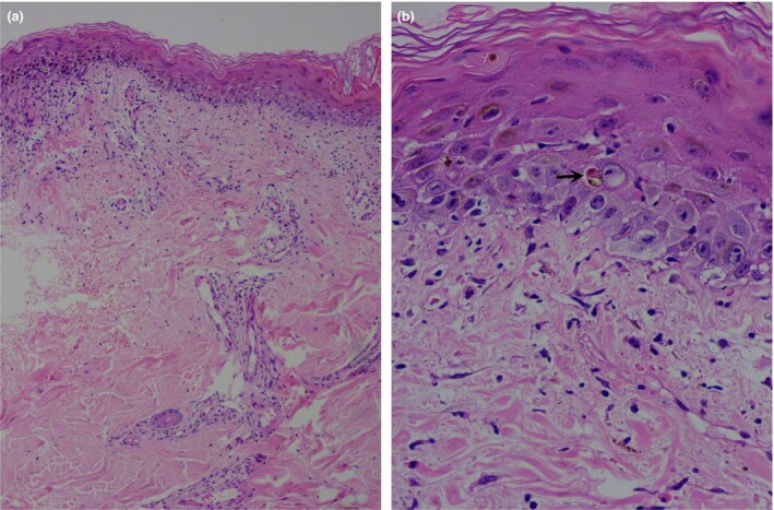
(a) Orthokeratosis with epidermal atrophy, scattered degenerated apoptotic keratinocytes, patchy areas of basal cell degeneration and interface dermatitis, perivascular and periadnexal inflammatory cell infiltrate, and extravasation of erythrocytes in the dermis; (b) apoptotic keratinocytes (arrow), upper dermal oedema and extravasation of erythrocytes. Haematoxylin and eosin, original magnification (a) × 50; (b) × 400.