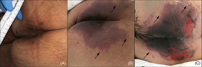
FIGURE 1.

Evolution of the patient's sacral/buttocks retiform purpuric patch during her COVID‐19 disease course. A, Baseline sacral skin photograph documented per standard protocol by the wound care team after consultation for intergluteal hyperpigmentation. The wound care team assessed that the hyperpigmentation was normal and there was no evidence of pressure injury or purpura. B, Retiform purpuric patch on Day 6 of hospitalization at time of initial dermatology evaluation; C, worsening of the purpuric patch with superficial ulceration on Day 12 of hospitalization at time of second dermatology evaluation. Arrows in panels B and C indicate branching retiform purpuric areas at the periphery of the wound edge, indicative of a thromboembolic process
