
FIGURE 2.
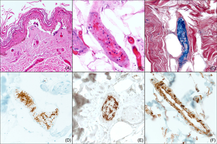
Histopathologic findings of a lesional biopsy specimen on hospitalization Day 6. Of note, biopsies from Days 6 and 12 revealed the same features: A, H&E sections reveal epidermal necrosis and papillary dermal vessels with intraluminal thrombi despite current therapeutic anticoagulation (×200). B, A deep dermal/subcutaneous vessel with extensive thrombosis admixed with inflammatory cell debris (×400). C, Phosphotungstic acid hematoxylin (PTAH) stain highlights fibrin‐rich thrombi staining blue within the vessels (×400). Platelet markers, D, CD61 (×400) and, E, CD41 (×400) reveal platelet aggregation within vessel lumina, with predominant aggregation at the vessel periphery. F, Complement split product C4d is diffusely present along the walls of vessel lumina (×400)