Figure 3.

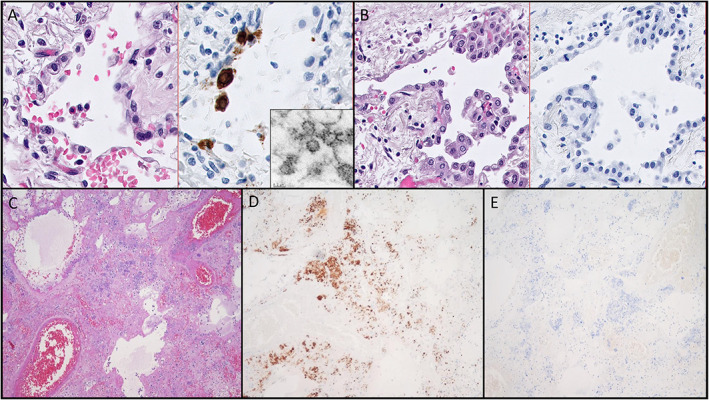
The nonspecific cytomorphology of SARS‐CoV‐2‐infected cells in human tissues and demonstration of cross‐reactivity with IHC reagents. (A) Hematoxylin and eosin (H&E)‐stained section (left) with paired IHC (right) of virus‐infected cells showing cytoplasmic vacuolization and reactive nuclear changes in infected cells (×600 magnification). Tissue dissected from this IHC‐positive region of the paraffin block demonstrates candidate virion‐like particles (inset, EM, ×80,000). (B) IHC of a group of cells from the same case (left H&E, right IHC) with identical cytomorphologic features which are negative for virus by IHC (×400 magnification). (C–E) Demonstration of nonspecific staining of bacteria by IHC in a case of necrotizing bacterial pneumonia and influenza from 2015. (C) H&E‐stained image of the region of positive staining (×100 magnification). (D) Positive staining of bacteria and bacteria‐laden macrophages by IHC (×200 magnification). (E) Negative ISH staining of the same region (×200 magnification). All magnifications represent total magnification.
