Figure 1.

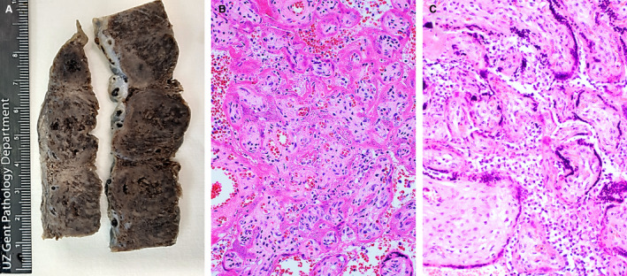
A, Macroscopic view of the placenta of case 1 showing large, irregular, solid areas with whitish discolouration. B, Haematoxylin and eosin (H&E)‐stained section of the placenta of case 1, showing necrotic syncytiotrophoblasts, collapse of the intervillous space, and some histiocytes in the remaining intervillous spaces. The villous stroma is well preserved. C, H&E‐stained section of the placenta of case 2, showing prominent histiocytic intervillositis. Syncytiotrophoblasts show pyknotic nuclei and focal loss of nuclear staining, indicating necrosis.
