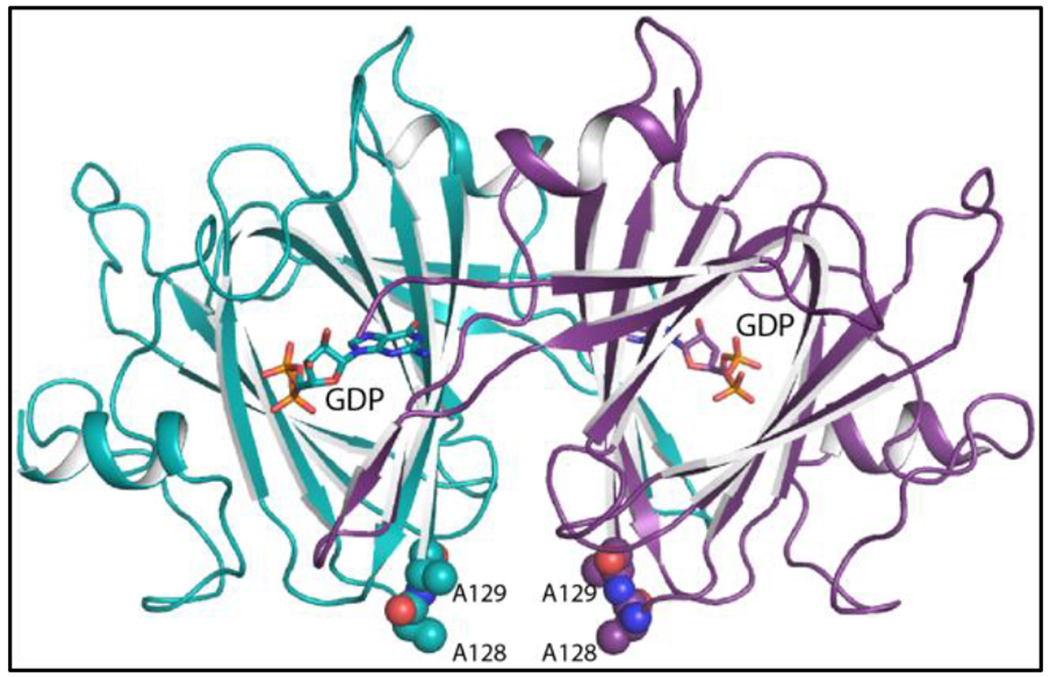
Figure 5.

Ribbon representation of the Cj1430 dimer. Subunits A and B are highlighted in purple and teal, respectively. The GDP ligands are displayed in stick representations. The positions the two site-directed mutations utilized to produce crystals with improved diffraction properties are depicted in sphere representations.
