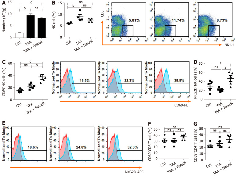
Figure 3.
Natural killer cells are activated in a Fasudil-treated liver fibrotic mouse model. After the last treatment, the mice were sacrificed and the intrahepatic lymphocytes were isolated. A: Absolute number of mononuclear cells in the liver from the normal control, thioacetamide (TAA)-induced, and TAA + Fasudil groups was assessed; B-G: Then the percentages and representative fluorescence-activated cell sorting (FACS) plots of natural killer (NK) cells among lymphocytes (B), CD69+ NK cells and representative FACS plots (C), quantification of NKG2D+ NK cells (D), representative FACS plots of NKG2D on NK cells (E), CD69+CD8+ T cells (F) and CD69+CD4+ T cells (G) in liver from the above three groups were analyzed by FACS. Data represent the mean ± SD from at least three independent experiments (n = 5/group). aP < 0.05, bP < 0.01, cP < 0.001.

