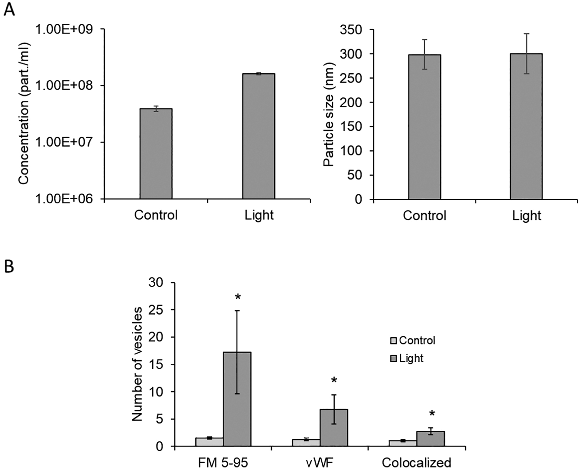
Figure 2. Red light induced exocytosis from pre-constricted murine facialis arteries.

Red light (670 nm, 6 J/cm2) induced dilation of blood vessel measured by pressure myography. The bath buffer was collected, centrifuged, and pelleted. (A): Extracellular vesicles in the reconstructed pellet were characterized using NanoSight particle tracking analyzer. (B): Endothelial origin was characterized by vesicle marker FM 5–95 and its co-localization with endothelial marker vWF. Values are mean±SE, *p<0.05 vs. Control.
