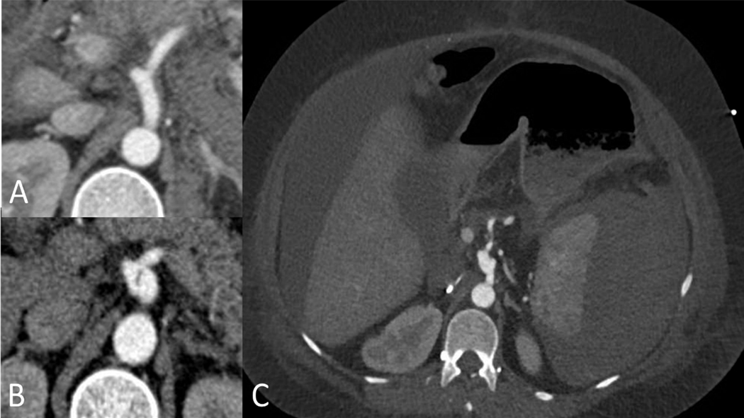
Figure 3.
Celiac artery dissection in 36 year old woman with vascular Ehlers-Danlos syndrome due to a splice site mutation (c.2337+2T>C/p.Gly762_Lys779del) managed medically. A. axial CT imaging obtained one month prior to the dissection when she presented with a spontaneous hemoperitoneum managed medically. B. Axial CT imaging demonstrating focal dissection and mild enlargement of the celiac artery. This remained unchanged on follow up imaging over the next 5 years. C. Axial imaging demonstrating a large spontaneous subcapsular splenic hematoma and hemoperitoneum. This occurred following a complicated hospitalization for perforated sigmoid diverticulitis requiring sigmoid resection, transverse colostomy and Hartmann’s pouch.

