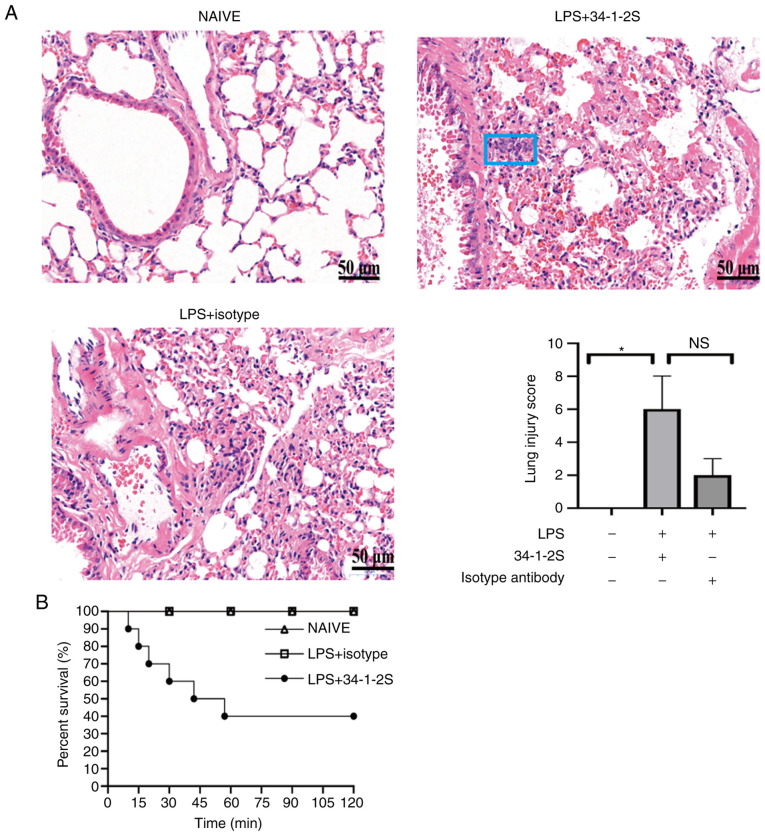
Figure 1.
Histological evidence and severity of antibody-mediated TRALI. (A) Hematoxylin and eosin stained lung histology sections photographed (magnification, ×400) and lung injury scores. Data are presented as the median and interquartile range (n=3/group). NS, no significance, *P<0.05, by Kruskal-Wallis test followed by Dunn's multiple comparisons test. The most representative of two replicate experiments, which were in good agreement, is shown. The blue rectangle indicates inflammatory cell accumulation. Scale bar, 50 µm. (B) Kaplan-Meier survival analysis of the treatment groups (n=10 mice/group). TRALI, transfusion-related acute lung injury; LPS, lipopolysaccharide; 34-1-2S, anti-MHC-I molecule.

