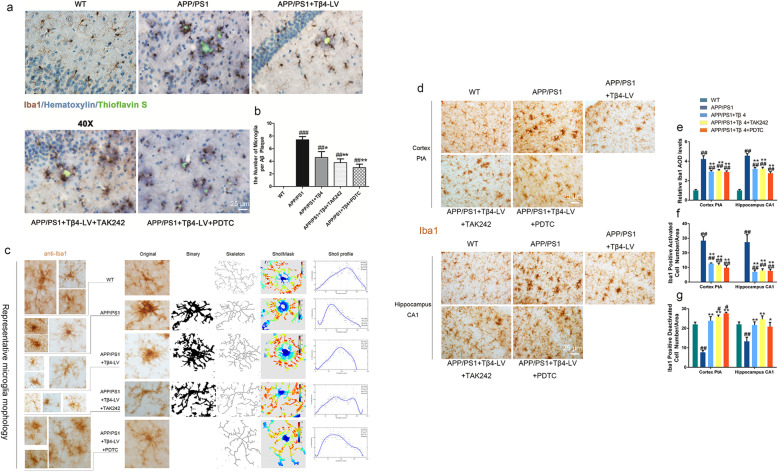
Fig. 4.
The effects of Tβ4 intervention and inflammatory pathway inhibition on microgliosis in the PtA and CA1 in groups of mice. a Representative images showed microglia infiltration implicated by Thioflavin S/IHC/Hematoxylin multiple staining in groups of mice. b Quantification of microglia surrounding per Aβ plaque. The data are presented as mean ± SEM (n = 4/group) in the different experimental groups. c Representative magnified profile of Iba1-labeled microglia morphology and process intersections by Sholl profile analysis in brain injection filed. d Representative images of Iba1-labeled microglia in PtA and CA1; quantification of e Iba1 immunopositivity, f number of Iba1-labeled activated and g deactivated microglia. The data are presented as mean ± SEM (n = 4/group) in the different experimental groups. #p < 0.05, ##p < 0.01 vs WT mice; *p < 0.05, **p < 0.01 vs APP/PS1 mice

