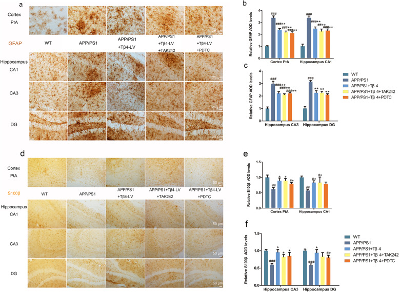
Fig. 6.
The effects of Tβ4 intervention and inflammatory pathway inhibition on astrogliosis and A2-phenotype of astrocyte in all groups of mice. a Representative images of GFAP-labeled astrocyte in PtA and hippocampus. b GFAP immunopositivity of PtA, CA1, and c CA3, DG region. The data are presented as mean ± SEM (n = 4/group) in the different experimental groups. d Representative photomicrographs of S100β positive cells. e S100β immunopositivity of PtA, CA1, and f CA3, DG region. The data are presented as mean ± SEM (n = 4/group) in the different experimental groups. #p < 0.05, ##p < 0.01, ###p < 0.001 vs WT mice; *p < 0.05, **p < 0.01, ***p < 0.001 vs APP/PS1 mice

