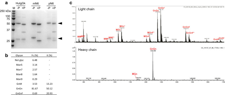
Fig. 4.
Glycosylation analysis of pN6. A PNGaseF assay where 1 µg each of PNGaseF-digested antibody (+P) was compared to undigested antibody (−P), including the positive control HuIgGk (human IgG1 kappa antibody, Sigma). Marker (M) is Precision Plus Protein™ All Blue Pre-stained Protein Standards. Heavy and light chains are indicated by black arrows. PNGase F enzyme visible at 36 kDa. B Percent abundance, derived from mass spectrometry, of various glycoforms in the heavy (Fc) and light (K) chains. C Mass spectra of pN6 heavy and light chain glycoforms. Purified proteins were analysed by digestion with trypsin followed by LC–ESI–MS. Glycopeptides from the kappa-chain variable region occurred as doubly charged ions, partly with ammonium

