Fig. 1.
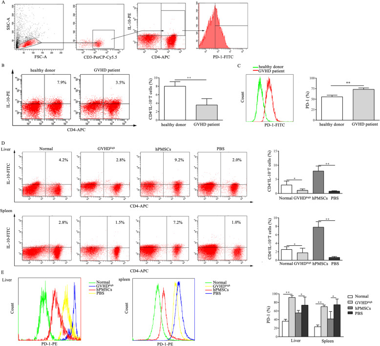
hPMSCs inhibit the expression of PD-1 in CD4+IL-10+ T cells during the development of GVHD. a Gating strategy. Forward scatter (FSC) and side scatter (SSC) gating were used to discriminate viable cells from cell debris. Within the lymphocyte gate, in cells obtained from GVHD patients and the GVHD mouse model, CD3 was used as a T cell marker, CD4+IL-10+ T cells were defined as those positive for CD4 and IL-10, and PD-1 expression in CD4+IL-10+ T cells was determined. b, c Dot plots of a representative experiment of CD4+IL-10+ T cells and the percentage of CD4+IL-10+ T cells and the statistical evaluation of the level of PD-1 in CD4+IL-10+ T cells obtained from GVHD patients and healthy donors (n = 7). d, e Representative dot plots and the percentage of CD4+IL-10+ T cells and the levels of PD-1 in CD4+IL-10+ T cells in the spleen and liver of GVHD mice in different groups (n = 5). The results were obtained from three independent experiments, *P < 0.05, **P < 0.01

