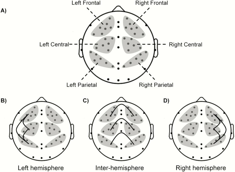
Figure 2.
(A) Topographical map showing EEG electrodes covering the frontal, central, and parietal regions of interest (ROIs; shaded gray regions). Overall, we investigated pairwise phase synchrony between six ROIs. (B) The three synchrony pairs among the three left-hemisphere ROIs, (C) six inter-hemispheric ROIs, and (D) three right-hemisphere ROIs.

