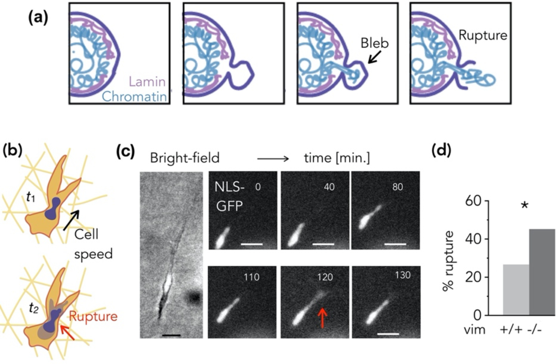
Figure 7.

(a) Schematic of nuclear envelope (NE) rupture. (b) Cells embedded in 3D collagen matrices exhibit spontaneous NE rupture. (c) Loss of NLS-GFP signal from the nucleus into the cytoplasm indicates NE rupture, and (d) cells lacking vimentin exhibit increased NE rupture.
