
Fig. 2. iRGD-liposome enhances homing and uptake of ASO in 4T1 and 22Rv1 tumor model.
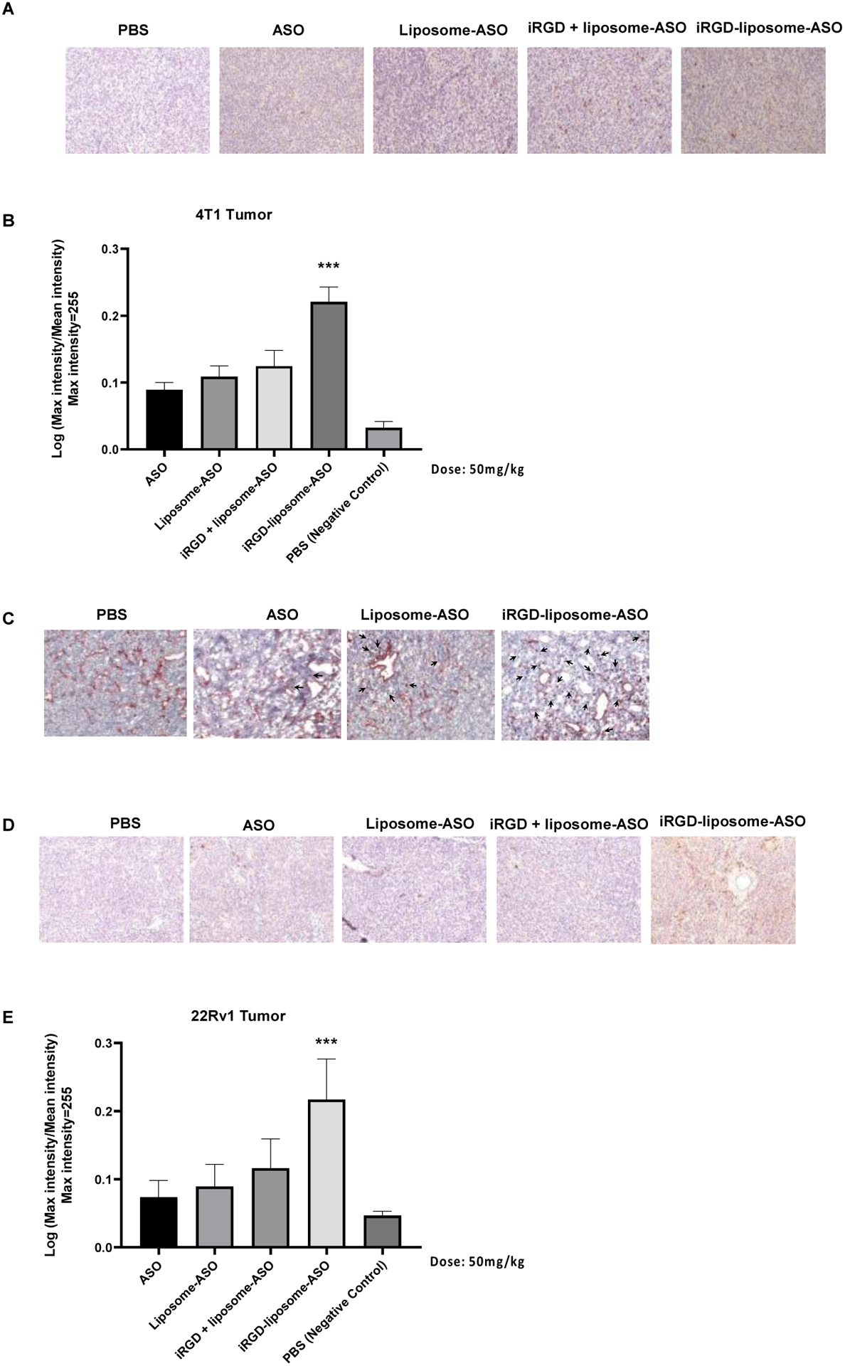
(A) Representative immunohistochemistry (IHC) sections and (B) quantification of ASO staining in 4T1 orthotopic breast tumors. The tumors were collected after 4-day homing by different delivery systems including PBS, free ASO, liposome-ASO, iRGD + liposome-ASO, and iRGD-liposome-ASO. The ASO dose was 50mg/kg. The density of ASO expression were analyzed by ImageJ. (C) IHC images of ASO accumulation in tumor stained with anti-ASO antibody for ASO (arrows) and anti-CD31 for blood vessels (red) after 2 h in vivo homing. (D) Representative immunohistochemistry (IHC) of ASO staining and (E) quantification of ASO in IHC sections from tumors of 22Rv1 tumor-bearing mice. The 22Rv1 tumor-bearing mice were treated by PBS, free ASO, liposome-ASO, iRGD + liposome-ASO, and iRGD-liposome-ASO, respectively. The ASO dose was 50mg/kg. The tumors were collected after 4-day homing by different delivery systems. All experiments were performed in three mice per group. The data are represented as mean ± standard deviation (SD). *P<0.05, ***P<0.001, ****P<0.0001 vs ASO. ASO: antisense oligonucleotide; AR: androgen receptor; The ASO dose was 50mg/kg. All experiments were performed in three mice per group.