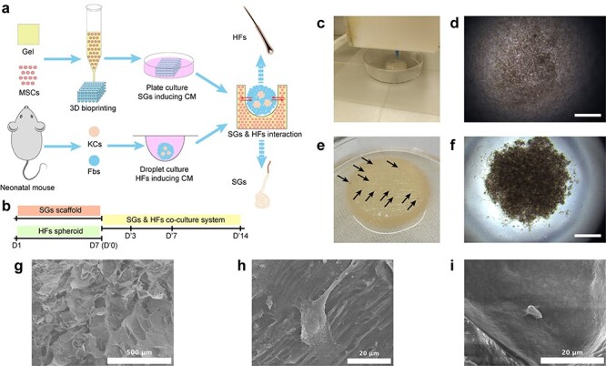
Figure 1.

Establishment of 3D skin constructs with multiple appendages. (a) Schematic diagram showing the procedure to establish 3D skin constructs in vitro. (b) Time points used in inducing SGs and HFs separately and SG–HF co-culture. (c) 3D bioprinting of SG scaffold. Brightfield imaging of HF spheroid in droplet culture 10 minutes (d) and 60 minutes (f) after seeding (scale bar, 300 μm). (e) Gross imaging of HF seeding on SG scaffold (black arrow shows gross view of HF spheroids seeded on SG scaffolds). (g) SEM shows the morphological structure of bioprinted AG constructs (scale bar, 500 μm). (h) SEM shows the morphology of MSCs in AG scaffold before SG induction (scale bar, 20 μm). (i) SEM shows the morphology of SG-like cells in SG scaffold after SG induction (scale bar, 20 μm). 3D three-dimensional, AG alginate–gelatin gel, CM culture medium, Fbs fibroblasts, HFs hair follicles, KCs keratinocytes, MSCs mesenchymal stem cells, SGs sweat glands, SEM scanning electron microscopy
