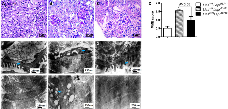
Figure 4.
Representative images of mesangial matrix expansion (MME), ultrastructural alterations of the podocytes and proximal tubule mitochondrial damage in 7-month-old mouse kidneys. (A) Lias+/+Leprdbdb/+, (B) Lias+/+Leprdbdb/db and (C) LiasH/HLeprdb/db mice, the sections stained with periodic acid–Schiff (PAS). Original magnification ×400. (D) Quantitative analysis of mesangial expansion in kidney glomerulus from the three strains. Data are expressed as mean±SEM. Podocyte foot process effacement was significantly increased in Lias+/+Leprdbdb/db mice (F) compared to Lias+/+Leprdbdb/+ mice (E). The increase was significantly improved in LiasH/HLeprdb/db mice (G). Original magnification, ×30,000. A large numbers of damaged mitochondria (blue arrowheads) in kidney proximal tubules of Lias+/+Leprdbdb/db mice (I), and the damaged mitochondria were seldom observed in proximal tubules of LiasH/HLeprdb/db mice (J) and Lias+/+Leprdbdb/+ mice (H). Original magnification ×30,000.

