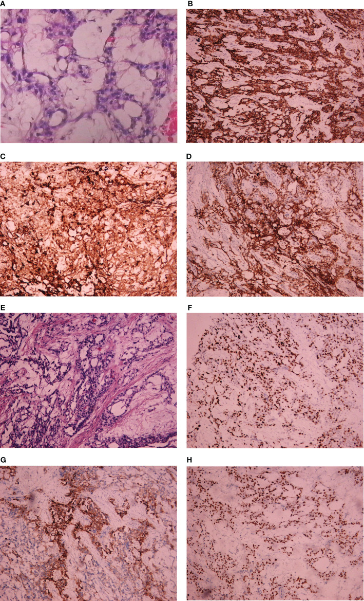
Figure 4.
Light microscopic appearance and immunohistochemical staining of YST originating from the broad ligament. (A) Yolk sac tumor (H and E, 10 × 40); (B) AE1/AE3+; (C) AFP++; (D) GPC-3(++); (E) Yolk sac tumor (H and E, 10 × 40); (F) Ki-67 positivity rate of approximately 70%; (G) PLAP negative; (H) SALL4 +++.

