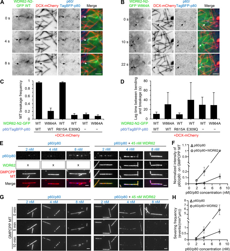
Figure 6.
WDR62 recruits katanin to sever curved MTs in cells and GMPCPP-MTs in vitro.(A and B) Time-lapse images of live MRC5 cells expressing GFP-tagged WT WDR62 N2 fragment (A) or its W864A mutant (B) together with DCX-mCherry and p60/TagBFP-p80. Arrows showing that p60/TagBFP-p80 was recruited to curved MT lattices by WDR62 N2 WT-GFP (A), but not by WDR62 N2 W864A-GFP (B), and that breakage occurred in the former but not the latter case. DCX-mCherry was used to distinguish between straight and curved MT lattices. (C) Quantification of MT breakage frequency in MRC5 cells expressing indicated constructs. n = 3 experiments; 90–120 curved MTs were measured. (D) Quantification of lag time between appearance of bending and breakage in MRC5 cells expressing indicated constructs. n = 90–100 breakage events for WDR62 N2 WT-GFP cotransfected with p60/TagBFP-p80 or p60/TagBFP-p80 R615A; n = 10–30 for other conditions. (E and F) Images and quantification of p60/SNAP-p80 (blue) intensity on GMPCPP-stabilized MTs (red) at various concentrations (2 nM, 4 nM, and 8 nM) in the absence (indicated with "x" in the left panels) or presence of 45 nM WDR62-GFP (green). The values were normalized to the intensity of 8 nM p60/SNAP-p80 in the presence of 45 nM WDR62-GFP. n = 40–60 MTs from three experiments. (G and H) Images and quantification of MT-severing frequency of p60/SNAP-p80 (2 nM, 4 nM, and 8 nM) in the absence or presence of 45 nM WDR62-GFP. n = 3 experiments; 40–60 MTs were measured. Scale bars, 2 µm. Data represent mean ± SD.

