Figure 7.
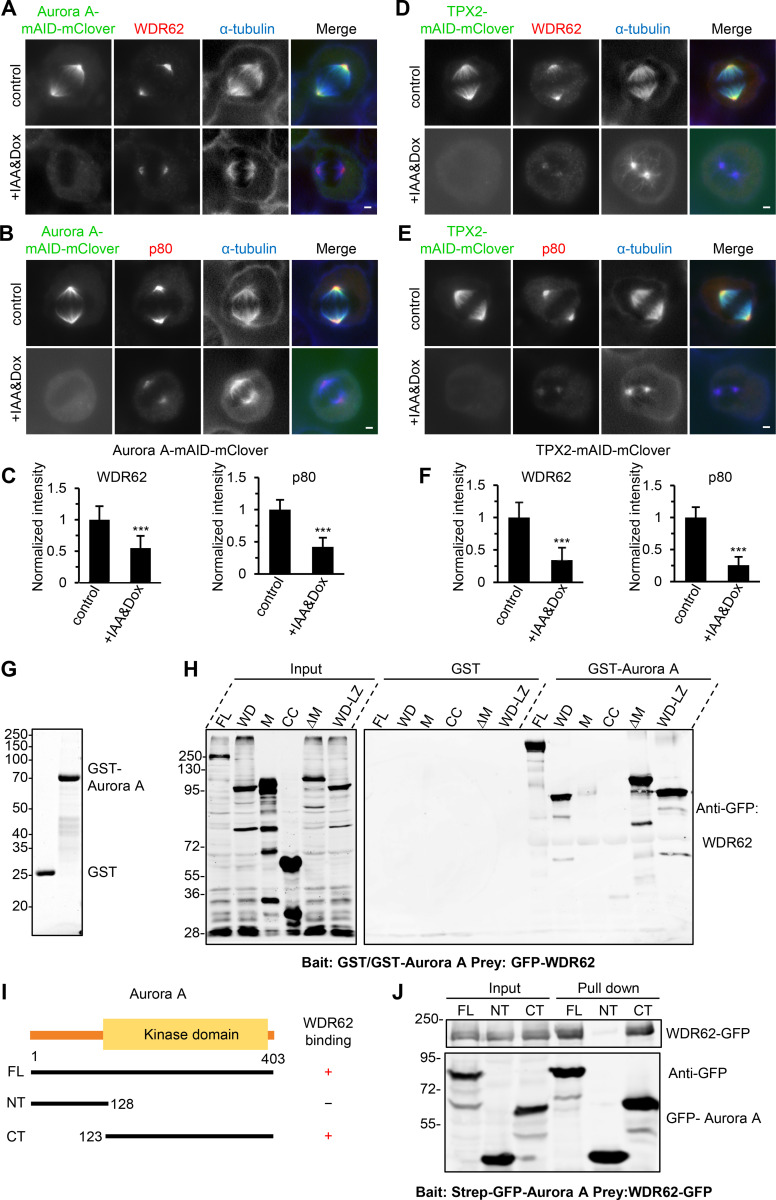
Validation of a TPX2–Aurora A–WDR62–katanin axis in cells.(A–C) Immunofluorescence staining and quantification of WDR62 and katanin p80 intensities at spindle poles in Aurora A–mAID-mClover knock-in HeLa cells without or with doxycycline (Dox) and IAA treatment. For WDR62 intensity, control, n = 102 spindle poles; doxycycline and IAA treatment, n = 108; for p80 intensity, both control and doxycycline and IAA treatment, n = 104. (D–F) Immunofluorescence staining and quantification of WDR62 and katanin p80 intensities at spindle poles in TPX2-mAID-mClover knock-in HeLa cells without or with doxycycline and IAA treatment. For all conditions, n = 100 spindle poles. (G) Coomassie blue–stained gel with GST and GST–Aurora A purified from E. coli and used for the GST pull-down assays in H. (H) GST or GST–Aurora A pull-down assays with extracts of HEK293T cells expressing GFP-tagged WDR62 FL or its indicated fragments analyzed by Western blotting with GFP antibody. M, middle region; CC, coiled-coil; LZ, leucine zipper. (I) Schematic overview of the domain organization of Aurora A and the deletion mutants and summary of their interactions with WDR62. (J) StrepTactin pull-down assays with extracts of HEK293T cells expressing Strep-GFP–tagged Aurora A FL, N-terminal part (NT), or C-terminal kinase domain (CT; bait) together with WDR62-GFP (prey) analyzed by Western blotting with GFP antibody. Scale bars, 2 µm. Data represent mean ± SD. ***, P < 0.001; two-tailed t test.

