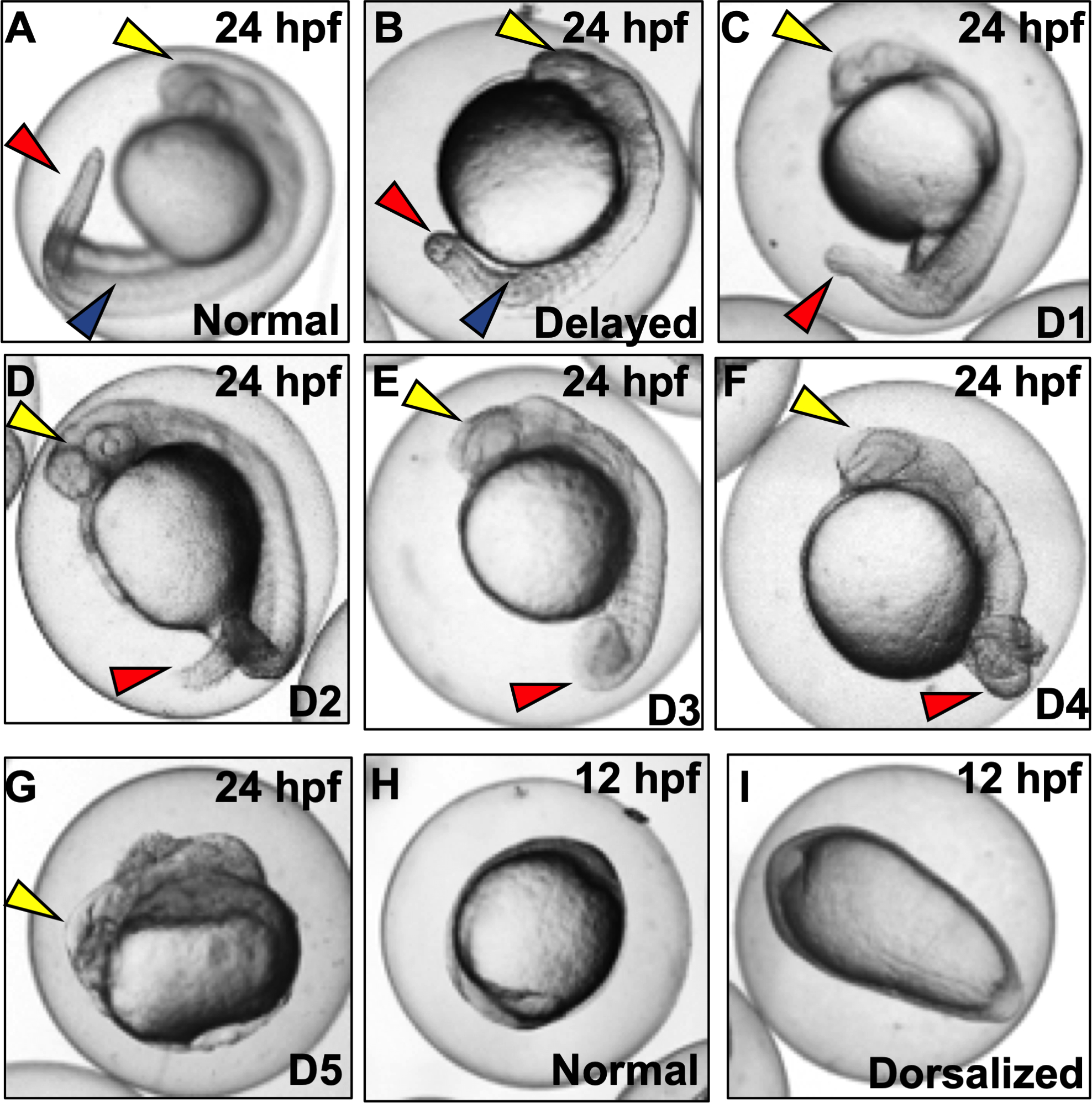
Figure 1. Representative images of dorsalization phenotypes (D1-D5) following exposures to vehicle (0.1% DMSO) or DMP (0.078–0.625 μM) with exposures initiated at 0.75 hpf (Basic Protocol 1).

(A) Normal embryo at the prim-5 stage. Yellow arrow denotes head; blue arrow denotes somites; and red arrow denotes tail. (B) Embryo delayed in development, with lack of development of eye/otic vesicle (yellow arrow) and somite numbers (blue arrow) compared to normal embryos. (C-D) Class 1–4 dorsalized embryos with red arrows representing loss of ventral tail fin and (E and F) gradual coiling of the tail and trunk area. (G) Class 5 dorsalization with ill-developed head (yellow arrow) but no development of trunk and tail tissues. (H and I) Normal and dorsalized (ovoid) embryos at 12 hpf.
