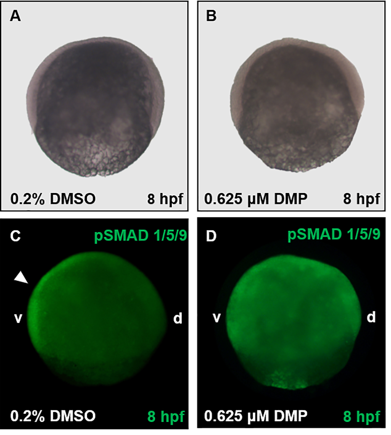
Figure 4. Representative images following pSMAD 1/5/9 localization (Basic Protocol 2).

Embryos were exposed to either vehicle (0.2% DMSO) (A, C) or 0.625 μM DMP (B, D) from 0.75 hpf and fixed in 4% PFA at 8 hpf. Embryos within Panels A and B were imaged using brightfield microscopy, and embryos within Panels C and D imaged using fluorescence microscopy. Within control embryos (C), pSMAD 1/5/9 staining is localized along the ventral (v) side of the embryo (white arrowhead), with lack of staining along the dorsal (d) side. Contrary to vehicle controls (C), the gradient of pSMAD 1/5/9 is disrupted by 0.625 μM DMP and diffused throughout the embryo (D).
