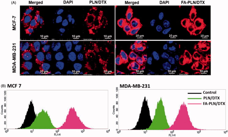
Figure 2.
(A) In vitro cellular uptake of PLN/DTX and FA-PLN/DTX in MCF-7 and MDA-MB-231 cancer cells. Cellular internalization was observed using a confocal laser scanning microscope (CLSM). Rhodamine-B was used as a fluorescent probe and nuclei were stained with DAPI. (B) Flow cytotmeter based cellular uptake of PLN/DTX and FA-PLN/DTX in MCF-7 and MDA-MB-231 cancer cells.

