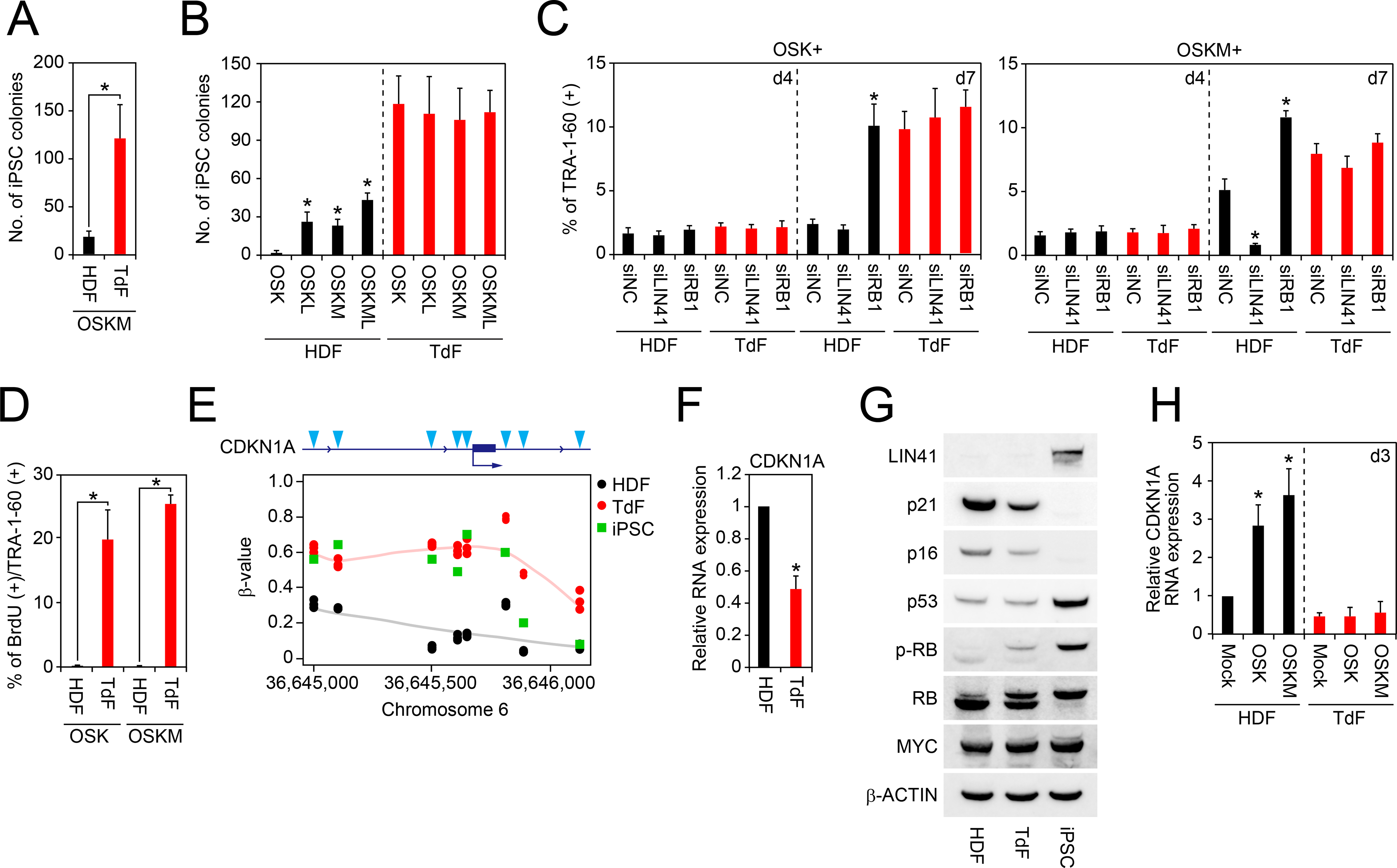
Figure 6: Immortalization Bypasses the Proliferation Pause.

A, The number of iPSC colonies from HDFs and TdFs. Shown are number of iPSC colonies on day 24 derived from 5 × 10⁴ cells of OSKM-transduced HDFs and TdFs. *p<0.05 by unpaired t-test (n=3).
B, Effects of exogenous MYC and LIN41 on iPSC generation from HDFs or TdFs. Shown are the number of iPSC colonies from HDFs or TdFs on day 24 post-transduction of OSK, OSKL, OSKM or OSKML. *p<0.05 by Dunnett’s test (n=3).
C, Effects of LIN41 or RB1 knockdown on the TRA-1–60 (+) cell proportion from HDFs or TdFs. Shown are the effects of LIN41 or RB1 knockdown by siRNA transfection on OSK or OSKM-induced TRA-1–60 (+) cell proportion from HDFs or TdFs on days 4 and 7. *p<0.05 by Dunnett’s test (n=3).
D, BrdU incorporation of newly converted TRA-1–60 (+) cells from TdFs or HDFs. Shown are the percentage of the BrdU incorporation of TRA-1–60 (+) cells from HDFs or TdFs on day 4 post-transduction of OSK or OSKM. *p<0.05 by unpaired t-test (n=3).
E, DNA methylation status at upstream region of CDKN1A gene. Shown are β-value of DNA methylation in each CpG site (blue arrowheads) at the upstream region of CDKN1A gene in HDFs (black), TdFs (red) and iPSCs (green) analyzed by Infinium. n=3.
F, Expression of CDKN1A in HDFs and TdFs. Shown is the relative expression of CDKN1A in HDFs and TdFs analyzed by qRT-PCR. *p<0.05 vs. HDFs by unpaired t-test (n=3).
G, Expression of cell-cycle-related proteins in HDFs, TdFs and iPSCs. Western blot showing expression of LIN41, p21, p16, p53, MYC and β-ACTIN proteins in HDFs, TdFs and iPSCs.
H, No induction of CDKN1A by OSKM in TdFs. Shown is the relative expression of CDKN1A mRNA in HDFs (black) or TdFs (red) on day 3 post-transduction of empty vector (Mock), OSK or OSKM. *p<0.05 by Dunnett’s test (n=3).
