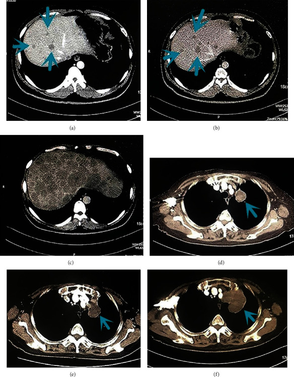
Figure 1.

Computed tomographic scans before baseline (a), at baseline about 6-8 weeks later (b), and during PD-1 and programmed death ligand 1 (PD-L1) inhibitor therapy 6-8 weeks later (c) in a man in his mid-40s with stage IV (mediastinal lymph nodes, right adrenal gland, and liver metastases) non-small-cell lung cancer treated with anti-PD-1 and anti-PD-L1 therapy in the sixth line. After 2 administrations, there was evidence of significant liver lesion progression. Arrowheads show liver lesions before and during anti-PD-1+anti-PD-L1 treatment. Computed tomographic scans before baseline (d), at baseline about 6-8 weeks later (e), and during programmed death 1 (PD-1) inhibitor therapy 6-8 weeks later (f) in a woman in her mid-70s with stage IV (hilar and mediastinal lymph nodes and jejunum metastases) EGFR L858R missense mutation and TP53 mutation lung adenocarcinoma treated with anti-PD-1 therapy in the third line. After 2 administrations, there was evidence of significant lung progression. Arrowheads show lung lesions before and during anti-PD-1 treatment.
