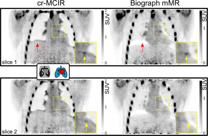
Fig. 4.
Result of the PET cr-MCIR application to patient 1. An enlarged ROI is marked by the dashed yellow square. Top, coronal slice 1; bottom, coronal slice 2. Left, cr-MCIR. Right, Biograph mMR vendor reconstruction. Uptake in the left coronary artery is highlighted by yellow arrows, and red arrows indicate artefacts due to an AC mismatch. The cr-MCIR shows reduced AC mismatch compared to the Biograph mMR images. For the vendor reconstruction, the strong AC data mismatch due to physiological motion means the uptake in the coronary plaque is not visible anymore in the second slice (bottom row). Scanner and STIR reconstructions required a different window setting for comparable contrast in the coronary uptake

