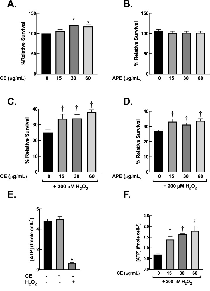
Figure 4.
Centella extracts prevent H2O2-induced cytotoxicity in dermal fibroblasts. (A) and (B) CE and APE at 15–60 µg/mL did not cause toxicity in dermal fibroblasts. BJ cells were incubated with CE or APE (15–60 µg/mL) for 24 h. (C) and (D) CE and APE inhibit oxidative damage from H2O2 exposure. BJ cells were pre-incubated with CE or APE (15–60 µg/mL) for 24 h then exposed to H2O2 (200 µM) for 1 h. The cell viability was evaluated with MTT assay immediately after the treatments. (E) and (F) CE inhibit depletion of intracellular ATP following H2O2 exposure. The protocol for treatment was as (A) and (C). The amounts of ATP were measured immediately after treatments (n = 3; mean ± SEM; *P < 0.05 vs untreated control; †P < 0.05 vs H2O2-treated cells).

