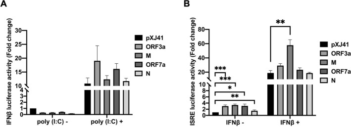
Figure 2.
Cellular IFN response mediated by SARS-CoV-2 proteins in HeLa cells. Cells were co-transfected with pIFN-β-Luc (0.5 μg) (A), or pISRE-Luc (0.5 μg) (B), along with pRL-TK (0.05 μg) and each (0.5 μg) of indicated SARS-CoV-2 genes. At 24 h post-transfection, the cells were transfected again with 0.5 μg of poly(I:C) for stimulation for 16 h (A), or incubated with IFN-β (1000 UI/ml) for 6 h (B). Cell lysates were then prepared for luciferase assays using the Dual Luciferase assay system according to the manufacture’s instruction (Promega). Relative luciferase activities were obtained by normalizing the firefly luciferase to Renilla luciferase activities. Values of the relative luciferase activity in the pXJ41 control group were set as 1, and the values for individual viral proteins were normalized using that of the pXJ41 control. Error bars mean ± standard deviation (s.d.). (n = 3). *P < 0.05, **P < 0.01, ***P < 0.001.

