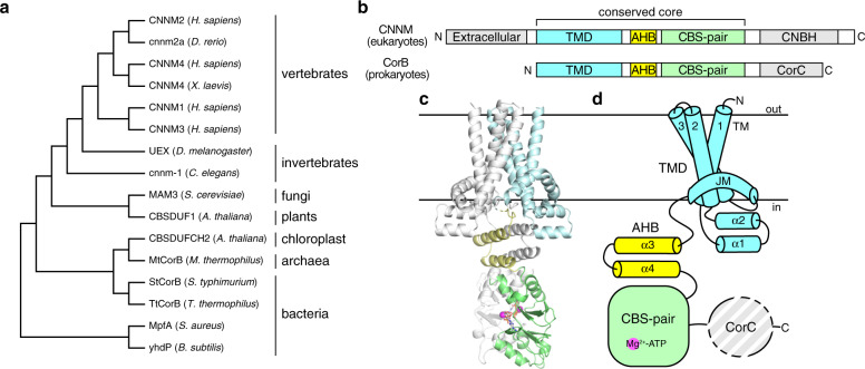
Fig. 1. Overall structure and domain organization of CNNM/CorB Mg2+ transporters.
a Phylogenetic analysis of representative CNNM/CorB orthologs generated using the neighbor-joining method. b Domain organization of eukaryotic CNNM and prokaryotic CorB. TMD transmembrane domain, AHB acidic helical bundle, CNBH cyclic nucleotide-binding homology domain, CorC cobalt resistance C domain. c Crystal structure of the Mg2+-ATP-bound MtCorB without the C-terminal CorC domain as a homodimer. One chain is colored by domains. d Topology of a MtCorB monomer showing the transmembrane and juxtamembrane helices of the TMD (residues 1–154, cyan), the two helices of the AHB (residues 166–199, yellow), the Mg2+-ATP-binding CBS-pair domain (residues 200–324, green), and the CorC domain (residues 325–426, grey).

