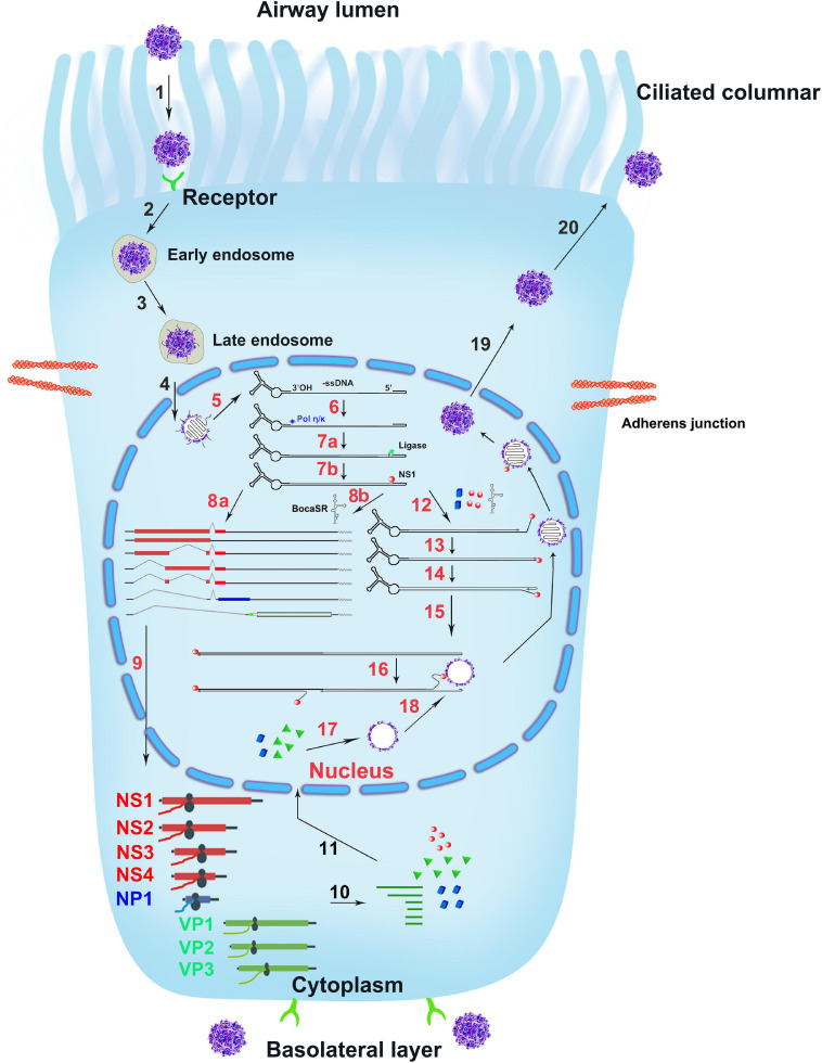
FIGURE 6.
The infection life cycle of HBoV1. A ciliated airway epithelial cell is depicted with diagrams of the cilia and junction molecules. HBoV1 enters the cells through binding to an unknown viral receptor, which is expressed on both the apical (ciliated) and the basal cells as indicated, and through receptor-mediated endocytosis, followed by intracellular trafficking (Steps 1–3). The virus escapes from the late endosome and enters the nucleus (Step 4). In the nucleus, the uncoated ssDNA viral genome is converted to replicative form dsDNA that expresses viral NS proteins and BocaSR (Steps 5–8). The viral DNA further replicates in the nucleus (Steps 12–16) and expresses both viral NS and capsid proteins (Steps 9–11), followed by genome packaging into empty capsid (Steps 16–18). Lastly, the matured virus egresses out of the infected cells (Steps 19, 20). The HBoV1 infection cycle in the ciliated epithelial cell is illustrated based on the studies on HBoV1 and references from other parvoviruses, which are explained in the text.

