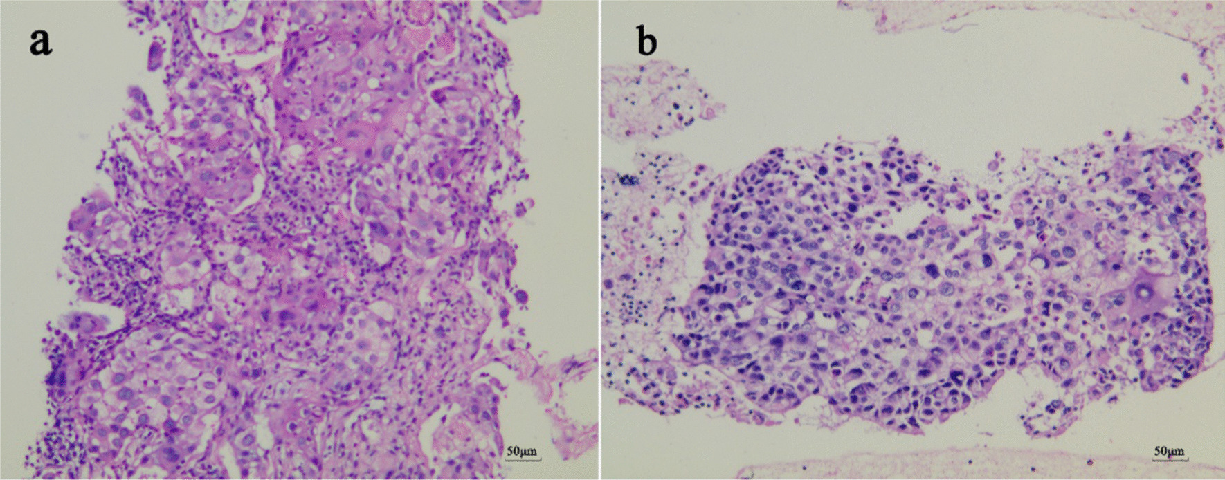
Fig. 3.

Hematoxylin and eosin sections (original magnification ×200). Slices with H&E staining were observed under the NIKON TE2000-U microscope(10×eyepiece, 20×objective lens). Photos were captured through a CCD digital camera involved in NIKON TE2000-U microscopic platform. Typical photos were recorded by NIKON NIS-Elements software V4.2. a CT-guided percutaneous lung biopsy. The tissue cut by fine needle was composed of atypical epithelioid neoplasm cells organized in a squamous carcinoma pattern. b EBUS-TBNA. Squamous cell clusters mixed in the mediastinal lymph node tissue of the N7 group aspirated by the fine needle
