. 2021 May 7;17(3):400–408. doi: 10.3988/jcn.2021.17.3.400

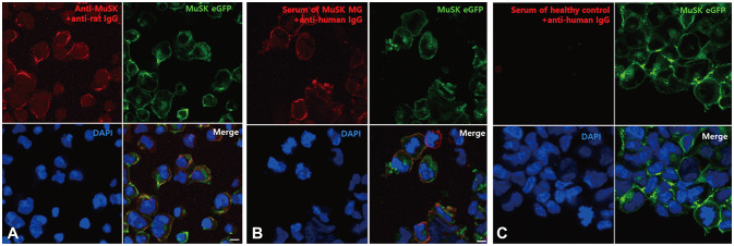
Fig. 2. CBA to detect anti-MuSK Abs. When HEK293 cells expressing MuSK were incubated with (A) commercially available polyclonal anti-MuSK Abs or (B) sera from patients with MuSK-Ab-positive MG, anti-IgG antibody binding (red fluorescence) was observed along the cell surface, indicating the binding of MuSK Abs to MuSK. This colocalized with GFP-labeled MuSK expressed on the surface of HEK293 cells (green fluorescence). In contrast, (C) anti-human IgG antibody binding (red fluorescence) was not observed after adding the sera from the healthy control. Scale bar: 10 µm. CBA: cell-based assay, DAPI: 4′,6-diamidino-2-phenylindole, eGFP: enhanced green fluorescent protein, HEK293: human embryonic kidney 293, MG: myasthenia gravis, MuSK Abs: antibodies against muscle-specific tyrosine kinase.

Fig. 2